
购物车0
产品总数:61756


| 中文名称 |
Mad1 (6-21)
|
|---|---|
| 英文名称 |
Mad1 (6-21)
|
| 英文别名 |
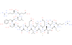
Mad1 (6-21);L-Arginine, L-arginyl-L-methionyl-L-asparaginyl-L-isoleucyl-L-glutaminyl-L-methionyl-L-leucyl-L-leucyl-L-α-glutamyl-L-alanyl-L-alanyl-L-α-aspartyl-L-tyrosyl-L-leucyl-L-α-glutamyl- (9CI)
|
| Cas No. |
880150-82-3
|
| 分子式 |
C84H140N24O26S2
|
| 分子量 |
1966.29
|
| 包装储存 |
Sealed storage, away from moisture and light, under nitrogenPowder -80°C 2 years;-20°C 1 ye
|
| 详情描述 |
Mad1 (6-21) 是 Mad1 蛋白的 6-21 片段。Mad1 (6-21) 与哺乳动物 Sin3A PAH2 结合,Kd 为 ~29 nM。
|
| 产品详情 |
Mad1 (6-21) 是 Mad1 蛋白的 6-21 片段。Mad1 (6-21) 与哺乳动物 Sin3A PAH2 结合,Kd 为 ~29 nM。
|
|---|---|
| 生物活性 |
Mad1 (6-21) is the 6-21 fragment of Mad1 protein. Mad1 (6-21) binds to mammalian Sin3A PAH2 with a K d of ~29 nM.
|
| 性状 |
Solid
|
| 体外研究(In Vitro) |
The PAH2 domain of mSin3A adopts a left-handed, up-and-down, four-helix bundle structure with residues in all four helices as well as in the turn regions defining a compact structural domain with an extensive hydrophobic core. Helices α1 and α2 form a deep hydrophobic pocket, which constitutes the primary interaction surface for the Mad1 (6-21) peptide. The Mad1 (6-21) forms an amphipathic α helix in the complex and interacts with PAH2 mainly through the apolar surface of the helix. has not independently confirmed the accuracy of these methods. They are for reference only.
|
| 运输条件 |
Room temperature in continental US; may vary elsewhere.
|
| 储存方式 |
Sealed storage, away from moisture and light, under nitrogenPowder -80°C 2 years;-20°C 1 ye
|
| SequenceShortening |
RMNIQMLLEAADYLER
|
| Solvent&Solubility |
In Vitro:
H2O Peptide Solubility and Storage Guidelines:1. Calculate the length of the peptide.2. Calculate the overall charge of the entire peptide according to the following table: |
| 参考文献 |
[1]. K Brubaker, et al. Solution structure of the interacting domains of the Mad-Sin3 complex: implications for recruitment of a chromatin-modifying complex. Cell. 2000 Nov 10;103(4):655-65.
|
1:一般建议:溶解度为Medlife测试数据,可能与文献描述存在差异。这是由于生产工艺和批次不同产生的正常现象。为了使其更好的溶解,请用37℃加热试管并在超声波水浴中震动片刻。不同批次产品溶解度各有差异,仅做参考,具体以实验方案为准。
2:储存条件:粉末-20°C一般情况可以保存3年,溶于溶剂-80°C一般情况可以保存1年。不同产品及不同批次产品可能存在差异,请细致阅读产品信息,并辅助参考相关文献描述。
Copyright © 2025 陌孚医药 All rights reserved 未经授权禁止拷贝本站所有资料,如有违反,将追究法律责任。
 沪ICP备2023012080号 |
 ![]() 沪公网安备31011402010657号
沪公网安备31011402010657号
 扫码关注公众号
 产品使用指南
