After administration of 4 mg/kg Salinomycin (Sal), 8 mg/kg Salinomycin and 10 uL/g saline water for 6 weeks, the mice are sacrificed. The size of the liver tumors in the Salinomycin treatment groups diminishes compare with the control group. The mean diameter of the tumors decreases from 12.17 mm to 3.67 mm (p<0.05) and the mean volume (V=length×width×0.5) of the tumors decreases from 819 mm to 25.25 mm (p<0.05). Next, the tumors are harvested, followed by HE staining, immunohistochemistry, and TUNEL assays, to assess the anti-tumor activity of Salinomycin. HE staining shows that the structure of the liver cancer tissue:nuclei of different sizes, hepatic cord structure is destroyed. Immunohistochemistry shows that PCNA expression is lower after Salinomycin treatment. HE staining and TUNEL assays indicates the Salinomycin-treated groups has higher apoptosis rates than control. Furthermore, immunohistochemistry shows an increased Bax/Bcl-2 ratio after Salinomycin treatment. The protein expression of β-catenin decreases in the Salinomycin treatment groups compared with control.
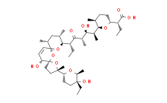
Salinomycin is a kind of monocarboxylic acid polyether type antibiotics, produced by the fermentation of Streptomyces albus, possesses a specific cyclic structure, and can form a complex compound with the pathogenic microorganisms and the extracellular cations of coccidian, especially K, Na, Rb, to alter the intracellular and extracellular ion concentrations.
Medlife has not independently confirmed the accuracy of these methods. They are for reference only.


 扫码关注公众号