
购物车0
产品总数:56415


| 中文名称 |
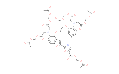
FURA-2AM
|
|---|---|
| 中文别名 |
荧光钙探针 Fura 2-AM;钙离子荧光探针 Fura 2-AM;AM荧光探针;钙离子荧光探针;钙荧光探针FURA-2, AM;FURA-2/AM荧光探针;FURA 2-AM SPECIAL PACKAGING
|
| 英文名称 |
Fura 2-AM
|
| 英文别名 |
Fura 2-AM;Fura-2 pentakis(acetoxymethyl) ester;2-(6-(Bis(2-((acetyloxy)methoxy)-2-oxoethyl)amino)-5-(2-(2-(bis(2-((acetylxoy)methoxy)-2-oxoethyl)amino)-5-methylphenoxy)ethoxy)-2-benzofuranyl)-5-oxazolecarboxylic acid (acetyloxy)methyl ester;Fura-2, AM;acetyloxymethyl 2-[5-[bis[2-(acetyloxymethoxy)-2-oxoethyl]amino]-4-[2-[2-[bis[2-(acetyloxymethoxy)-2-oxoethyl]amino]-5-methylphenoxy]ethoxy]-1-benzofuran-2-yl]-1,3-oxazole-5-carboxylate;Fura 2-AM Special Packaging;Fura-2-Acetoxymethyl ester;FURA 2;Fura 2am;Fura 2-AM 1mM;Fura-2(AM);l DMSO Solution;Fura?-AM;Fura 2 Am Ester;Fura-2 acetoxymethyl;fura 2-am yellow powder;Fura 2 acetoxymethyl ester;AM in Solution;AM, FOR FLUORESCENCE;FURA-2;FURA 2/AM;FURA 2/AM in Solution;FURA-2/AM, FOR FLUORESCENCE;FURA2/AM *1SET = 10x100UG**;Fura-2, AM *CAS 108964-32-5*;Fura-2 AM
|
| Cas No. |
108964-32-5
|
| 分子式 |
C44H47N3O24
|
| 分子量 |
1001.85
|
| 包装储存 |
-20°C, protect from light *该产品在溶液状态不稳定,建议您现用现配,即刻使用。 |
| 详情描述 |
108964-32-5,FURA-2AM,Medlife,上海现货。 Medlife,致力于提供高品质、高性价比小分子化合物的产品。 Medlife小分子化合物大量库存,提供超过2万种的抑制剂、激动剂、拮抗剂等产品,是药物及疾病研究的重要原料供应商。 荧光Ca2+指示剂,Fura-2 AM 是一种比率荧光 Ca2+ 指示剂。 查询关键词:“108964-32-5,FURA-2AM,PC15195,Medlife,上海现货”。 |
| 生物活性 |
Fura-2 AM is a high affinity, intracellular, UV light-excitable and ratiometric fluorescent Ca indicator. |
|---|---|
| 性状 |
Liquid |
| 体外研究(In Vitro) |
Guidelines (Following is our recommended protocol. This protocol only provides a guideline, and should be modified according to your specific needs). Medlife has not independently confirmed the accuracy of these methods. They are for reference only. |
| 运输条件 |
Room temperature or refrigerated transportation. |
| 储存方式 |
-20°C, protect from light *该产品在溶液状态不稳定,建议您现用现配,即刻使用。 |
| 参考文献 |
1:一般建议:溶解度为Medlife测试数据,可能与文献描述存在差异。这是由于生产工艺和批次不同产生的正常现象。为了使其更好的溶解,请用37℃加热试管并在超声波水浴中震动片刻。不同批次产品溶解度各有差异,仅做参考,具体以实验方案为准。
2:储存条件:粉末-20°C一般情况可以保存3年,溶于溶剂-80°C一般情况可以保存1年。不同产品及不同批次产品可能存在差异,请细致阅读产品信息,并辅助参考相关文献描述。
Copyright © 2025 陌孚医药 All rights reserved 未经授权禁止拷贝本站所有资料,如有违反,将追究法律责任。
 沪ICP备2023012080号 |
 ![]() 沪公网安备31011402010657号
沪公网安备31011402010657号
 扫码关注公众号
 产品使用指南
